Proceratium in China

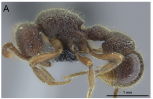
We have a new paper out in Zookeys (https://doi.org/10.3897/zookeys.770.24908) that revises the taxonomy of the very rare and cryptic ant genus Proceratium in China. We recognized 8 species from China and described 3 of them as new to science. The most spectacular species from Yunnan we named Proceratium shohei in honor of Dr. Shohei Suzuki (1979-2016), a marine scientist from OIST who lost his life in a tragic research diving accident. The study was led by our colleague Michael Staab, and included Paco and Cong from the lab, along with Zheng-Hui Xu from China.

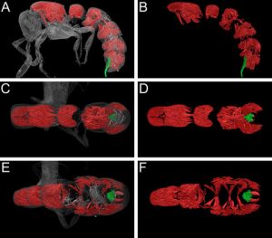
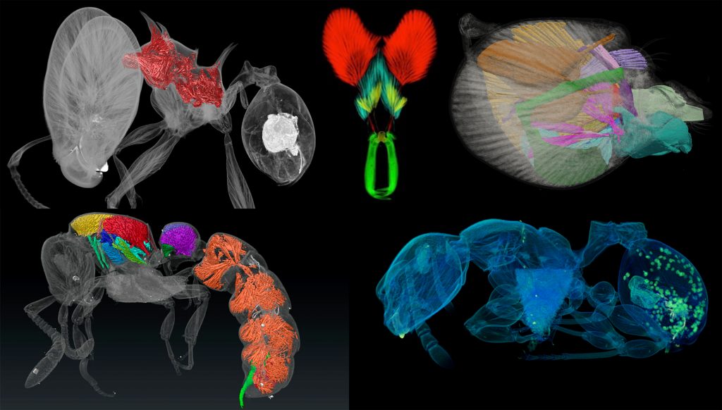
This study continues our lab’s line of research integrating x-ray microtomography (micro-CT) scanning, computer-based 3D reconstructions, and several downstream 3D data products (such as 3D surfaces and videos) into ant systematics. We used virtual 3D surface models based on micro-CT scans for in-depth comparative analyses of specimen morphology in order to overcome the difficulties of examining the rare and valuable physical material. Since these ants are extremely hairy, thus often very dirty, we basically “shaved” them virtually.


OIST minisymposium on using advanced imaging techniques to study evolution of ant phenotypes

Last week our lab hosted an OIST Mini Symposium titled “Advances in imaging, quantifying, and understanding the evolution of ant phenotypes” organized by Evan Economo and Francisco Hita Garcia. The aim of the symposium was to gather a small but selected group of leading researchers interested in the evolution of ant phenotypes with a strong focus on the use of x-ray microtomography (micro-CT). Our list of speakers covered experts in the fields of molecular and morphological systematics, anatomy, functional morphology, comparative morphology, adaptive trait evolution, reproductive biology, linear and geometric morphometrics, and paleontology. All invitees gave outstanding talks and presented published or ongoing research in great detail and with beautiful 2D or 3D illustrations and/or videos.
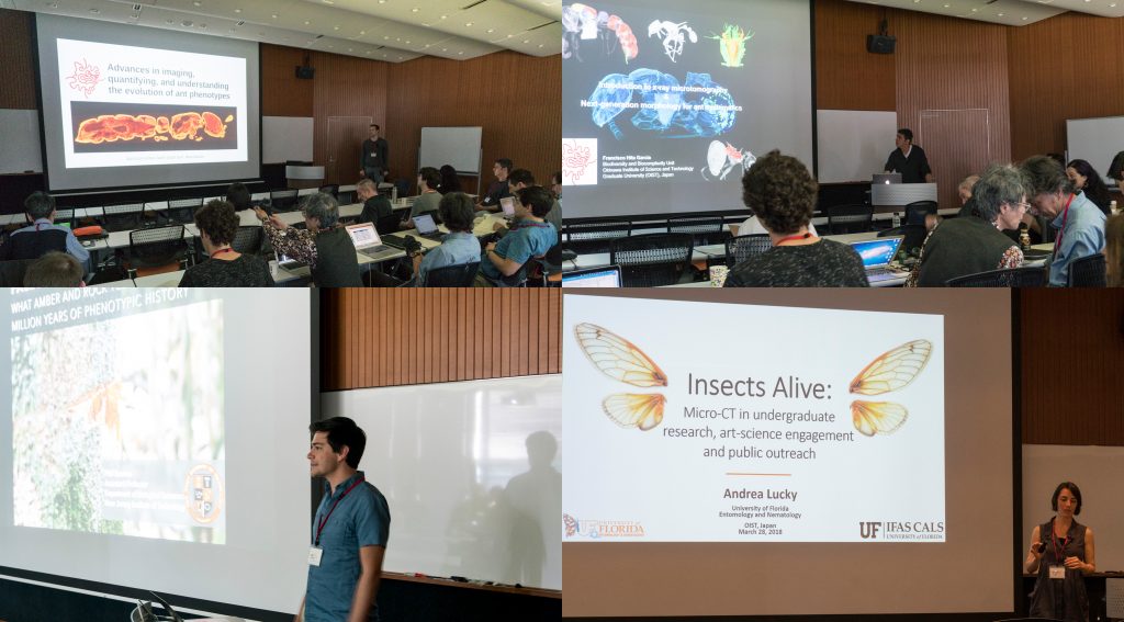
Some talks provided conceptual and technical backgrounds and perspectives of how to use micro-CT for ant morphology, how to better integrate next-generation phenomics into systematics, palaeontology, and evolutionary biology, and how to use micro-CT data and downstream 3D applications for education and public outreach.
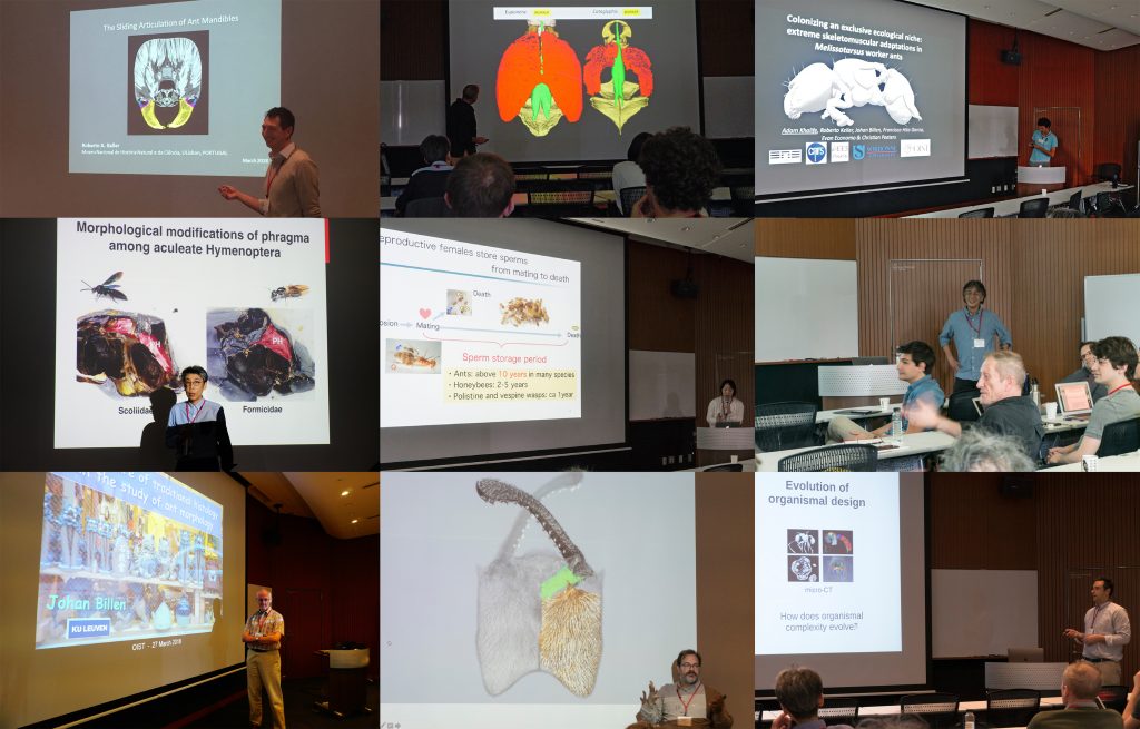
A strong focus of the symposium was the use of micro-CT for ant functional morphology, biomechanics, and the evolution of complex phenotypes. Some guests also showed recent advances in histology-based anatomy and reproductive biology, and shared ideas of how to combine traditional histology with modern 3D imaging technologies, such as micro-CT.
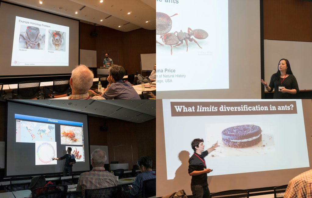
We also had a session focusing on the use of 2D linear and 3D geometric morphometrics and their application for ant phylogenetics, taxonomy, trait evolution, and more generally how to use large 3D phenotypical datasets to answer questions in evolutionary biology.
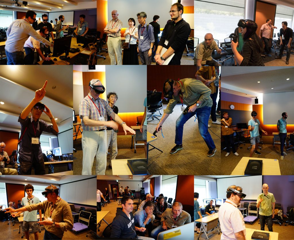
One afternoon was completely devoted to practical demonstrations of how to use 3D data. Our lab shared how we scan data post-processing, 3D virtual reconstructions, 3D animations, virtual/augmented reality, 3D printing. It was useful for sharing knowledge of methodology, and stimulating ideas for future directions and applications.
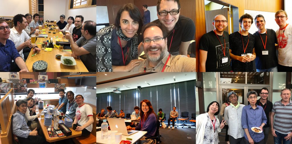
The three-day symposium provided ample opportunities for socializing and chatting about on-going and potential collaborations, discussions about methods and research results, as well as brainstorming about future directions for the field. At the same time our invitees got the chance to enjoy Japanese and Okinawan culture and cuisine and show off their Karaoke skills.

Invited speakers:
Phil Barden (New Jersey Institute of Technology)
Johan Billen (KU Leuven)
Benjamin Blanchard (U. Chicago and Field Museum)
Ayako Gotoh (Konan U.)
Yoshiaki Hashimoto (U. Hyogo, Museum of Nature and Human Activities, Hyogo)
Fuminori Ito (Kagawa U.)
Roberto Keller (Museu Nacional de História Natural e da Ciência)
Andrea Lucky (U. Florida)
Christian Peeters (U. Pierre et Marie Curie)
Shauna Price (Field Museum)
Andrew Suarez (U. Illinois)

Internal speakers:
Evan P. Economo
Georg Fischer
Nick Friedman
Francisco Hita Garcia
Adam Khalife (U. Pierre et Marie Curie and OIST)
New paper on 3D ant systematics

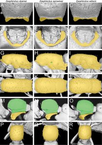
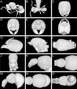
We have a new paper out today in ZooKeys revising the doryline genus Zasphinctus in the Afrotropical region. Led by Paco Hita Garcia, we do a deep dive into using microCT and 3D data for ant taxonomy. In previous recent papers, we provided 3D models and virtual type specimens to support taxonomic work. Here we go further and exploit more fully the power of micro-CT to discover and examine characters useful for systematics and the 3D representation of virtual specimens.

The three species are named after three important figures in biodiversity conservation. We named one after former US President Barack Obama, for his role in protecting natural areas. The species was found within a few kilometers of Obama’s father’s village in Kenya. The second species was named after E.O. Wilson, discovered from Gorongosa National Park, Mozambique, where he and his foundation have done a lot of work over recent years. The third species is named after Nigerian environmental activist Ken Saro-Wiwa.

Read the paper.

Interact with the 3D models on Sketchfab.

OIST media release.


New paper on evolution of ant spinescence in Pheidole

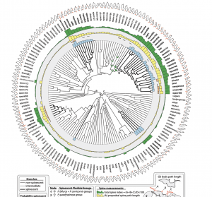
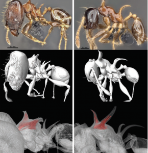
A new paper from the lab was published today in the Biological Journal of the Linnean Society focusing on aberrant spinescent phenotypes in Pheidole (including the famous dragon ants). We look at spinescence from a number of angles including phylogenetic, ecological, geographic, and 3D morphology. This study sheds light on the complexity of the issue of spine phenotype evolution. There are a number of open questions and some big mysteries. For starters, why the heck has spinescence evolved so many times in the Indo-Pacific, but no spiny Pheidole in New World? Check out the paper here!


HoloLens comes to the Economo Unit

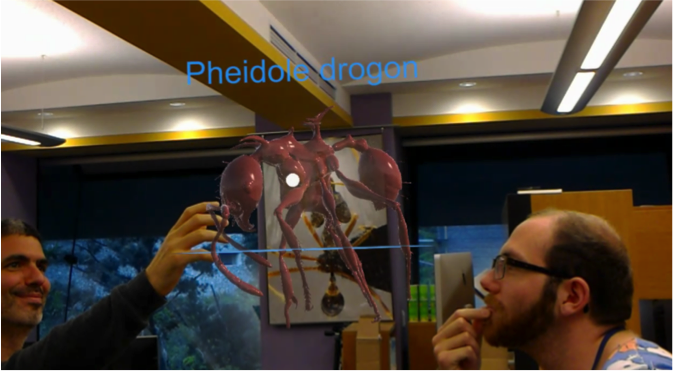
How would you like to be in the same room as an ant the size of a horse? In reality that’s a ridiculous idea, but in augmented reality anything is possible. This week, OIST’s Biodiversity and Biocomplexity Unit tested a new augmented reality device: the Microsoft HoloLens. Pavel Puchenkov of OIST’s Scientific Computing & Data Analysis Section designed a demonstration using a 3D-scanned model of the newly discovered ant species, Pheidole drogon. With the HoloLens, researchers were able to explore this species’ exceptionally spiny shape.

The view from the HoloLens. Paco Hita Garcia and Sam Ross can’t
actually see the ant, but it all looks very dramatic

While the applications available for the HoloLens are currently limited, the opportunities quickly become apparent. The ability to display holographic renderings of specimens in 3D has the potential to turn any room into a stunning museum exhibit. It’s clear that this technology has the potential to create or enrich experiences that we can use to communicate life’s diversity and evolutionary history.

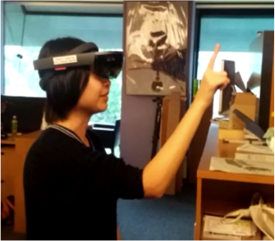
Ph.D. Student Yuka Suzuki interacting with objects in augmented reality.

Post written by Nicholas Friedman