OIST minisymposium on using advanced imaging techniques to study evolution of ant phenotypes

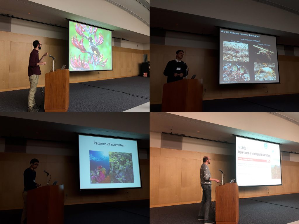
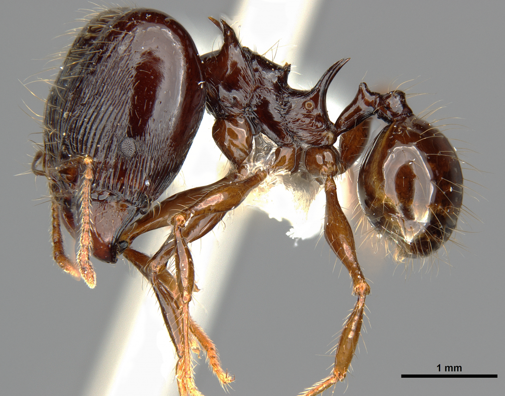
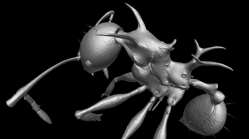
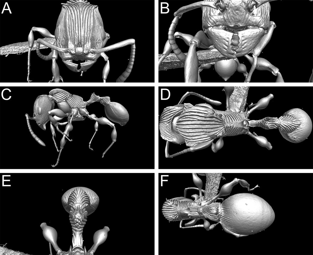
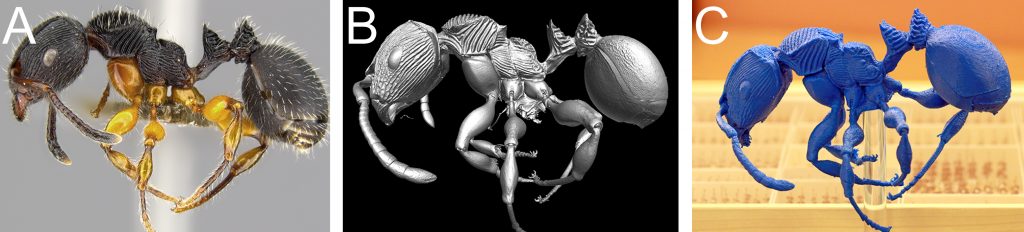
Last week our lab hosted an OIST Mini Symposium titled “Advances in imaging, quantifying, and understanding the evolution of ant phenotypes” organized by Evan Economo and Francisco Hita Garcia. The aim of the symposium was to gather a small but selected group of leading researchers interested in the evolution of ant phenotypes with a strong focus on the use of x-ray microtomography (micro-CT). Our list of speakers covered experts in the fields of molecular and morphological systematics, anatomy, functional morphology, comparative morphology, adaptive trait evolution, reproductive biology, linear and geometric morphometrics, and paleontology. All invitees gave outstanding talks and presented published or ongoing research in great detail and with beautiful 2D or 3D illustrations and/or videos.

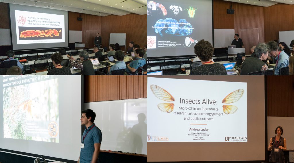
Some talks provided conceptual and technical backgrounds and perspectives of how to use micro-CT for ant morphology, how to better integrate next-generation phenomics into systematics, palaeontology, and evolutionary biology, and how to use micro-CT data and downstream 3D applications for education and public outreach.

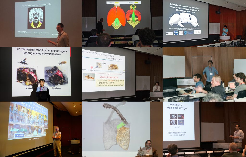
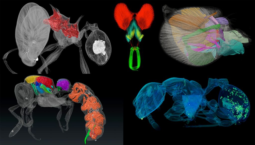
A strong focus of the symposium was the use of micro-CT for ant functional morphology, biomechanics, and the evolution of complex phenotypes. Some guests also showed recent advances in histology-based anatomy and reproductive biology, and shared ideas of how to combine traditional histology with modern 3D imaging technologies, such as micro-CT.

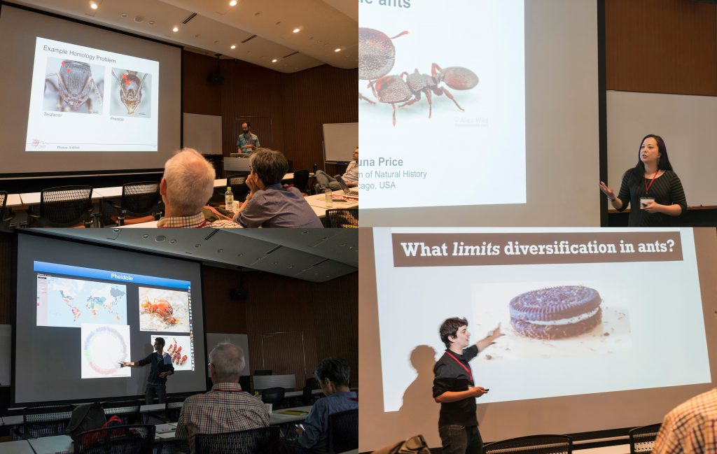
We also had a session focusing on the use of 2D linear and 3D geometric morphometrics and their application for ant phylogenetics, taxonomy, trait evolution, and more generally how to use large 3D phenotypical datasets to answer questions in evolutionary biology.

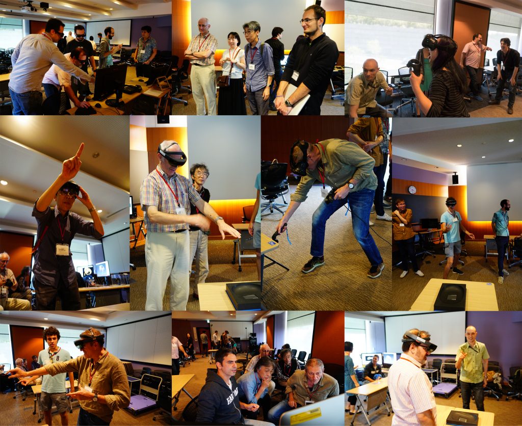
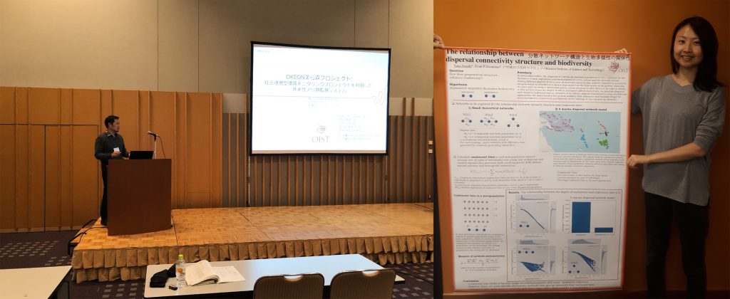
One afternoon was completely devoted to practical demonstrations of how to use 3D data. Our lab shared how we scan data post-processing, 3D virtual reconstructions, 3D animations, virtual/augmented reality, 3D printing. It was useful for sharing knowledge of methodology, and stimulating ideas for future directions and applications.

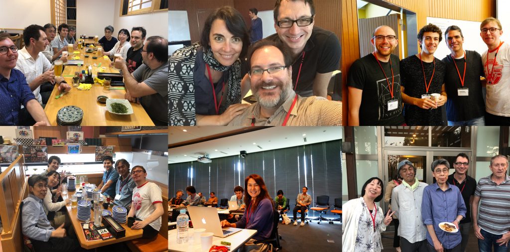
The three-day symposium provided ample opportunities for socializing and chatting about on-going and potential collaborations, discussions about methods and research results, as well as brainstorming about future directions for the field. At the same time our invitees got the chance to enjoy Japanese and Okinawan culture and cuisine and show off their Karaoke skills.

Invited speakers:
Phil Barden (New Jersey Institute of Technology)
Johan Billen (KU Leuven)
Benjamin Blanchard (U. Chicago and Field Museum)
Ayako Gotoh (Konan U.)
Yoshiaki Hashimoto (U. Hyogo, Museum of Nature and Human Activities, Hyogo)
Fuminori Ito (Kagawa U.)
Roberto Keller (Museu Nacional de História Natural e da Ciência)
Andrea Lucky (U. Florida)
Christian Peeters (U. Pierre et Marie Curie)
Shauna Price (Field Museum)
Andrew Suarez (U. Illinois)

Internal speakers:
Evan P. Economo
Georg Fischer
Nick Friedman
Francisco Hita Garcia
Adam Khalife (U. Pierre et Marie Curie and OIST)

ESJ 2018 in Sapporo

Several Arilab members attended the 65th Annual Meeting of the Ecological Society of Japan in Sapporo, Hokkeido, from March 14th to march 18th. It was a great opportunity for students, postdocs, and staff scientists to present Arilab research to a broader Japanese audience. At the same time, it was a good occasion for networking, chatting about potential future collaborations, and learn more about the research done in other labs throughout Japan.

Spearheaded by Nick Friedman and supported by Nao Takashina and Francisco Hita Garcia our lab organized a successful English-speaking symposium with the title “Biodiversity: linking biogeographic pattern and process”.

Masashi Yoshimura also gave an interesting about the OKEON Churamori Project and Yuka Suzuki successfully presented her poster showing her PhD project.

We also enjoyed the local Hokkeido cuisine, especially a visit to the Sapporo Bier Garten!

The dragon ant makes it into the Top10 New Species of 2017

Pheidole drogon major worker

Since 2008, the International Institute for Species Exploration in New York, USA publishes an annual Top 10 list of the new species discovered the past year. The annual list is released around May 23 to honor of the birthday of Carl Linnaeus, also known as the Father of Taxonomy.

Amidst a rat, a worm, a stingray, two plants and other arthropods, the dragon ant Pheidole drogon that was described last year by researchers in our lab made the cut to the 2017 Top 10.

Along with Pheidole viserion, Pheidole drogon – found in Papua New Guinea – owes its name to the dragon in the famed Game of Thrones book and TV series. The idea was inspired from the large spines on the back of the ant, which is revealed to be a location for muscle attachment to allow great strength in the head and mandibles.

The 2017 list of nominees also includes the bleeding ‘Bloodybone’ bush tomato, the spider Eriovixia gryffindori resembling the ‘Sorting Hat’ in the Harry Potter series, an amphibious centipede, a marine worm that look like a churro fried pastry, a South American plant which flower looks like a “Devil head”, a large spotted freshwater stingray, a millipede that continuously adds extra limbs throughout its lifetime, a vegetable-eating rat and finally a leaf-like katydid.

The institute’s international committee of taxonomists selects the Top 10 from among the approximately 18,000 new species named the previous year.

Pheidole drogon minor worker

Written by the OIST Media Section, edited by Julia Janicki.

New paper published: Patterns and processes in mountain ant metacommunities

Understanding the drivers of biodiversity patterns is always difficult due to the fact that multiple factors such as environmental gradient and spatial connectivity might contribute to the species distribution and community composition patterns simultaneously.

In a new paper just published in Ecography, we (Liu, Dudley, Xu and Economo) evaluate the effects of environmental gradients and spatial connectivity on ant taxonomic and phylogenetic diversity patterns along a 5000m elevational gradient within a complex mountainous landscape in Hengduan Mountains, a biodiversity hotspot in Southwest China.


We found that environmental gradients dominate variation in both alpha and beta diversity in this landscape, with alpha diversity strongly declining with elevation and beta diversity driven by elevational differences. We compared our system to predictions of a recent theoretical framework (Bertuzzo et al. 2016; PNAS) which synthesizes how aspects of landscape geomorphology may drive biodiversity patterns in idealized mountain landscapes. Our findings did not match the theory, we found alpha diversity is monotonically declining and within-band beta diversity is invariant with increasing elevation, but point toward ways to improve the theory. Taken together, our results show how elevation-driven environmental gradients, spatial factors, as well as landscape geomorphology together affect ant metacommunity structure in a complex mountainous landscape.

Original paper can be found here

New paper published: Potentials of micro CT for ant taxonomy

In the paper we explore the potential of x-ray micro computed tomography (μCT) for the field of ant taxonomy and use it intensively for the descriptions of two remarkable new species of the genus Terataner from Madagascar. In addition to the traditional way of presenting new species with stacked montage light photography, we also provide 3D models based on μCT data and make the whole 3D datasets available online through Dryad.

One important aspect of the study is to assess how μCT can improve collections-based research of ants and other insects. Our μCT-based 3D models can be virtually rotated, sectioned, measured, and dissected, thus allowing a wide range of analyses of the anatomy and morphology of the studied organisms. By generating and presenting virtual 3D models of ants (or other animals) we support the establishment of virtual natural history collections that permit rapid and free access to anatomically correct and permanent digital reconstructions or avatars of physical specimens. Another great advantage is of the technology is the ability to print physical models of the scanned specimens, which can be used for a variety of research, museum, educational, and outreach purposes.